Overactivation of histone deacetylases and EZH2 in Wilms tumorigenesis


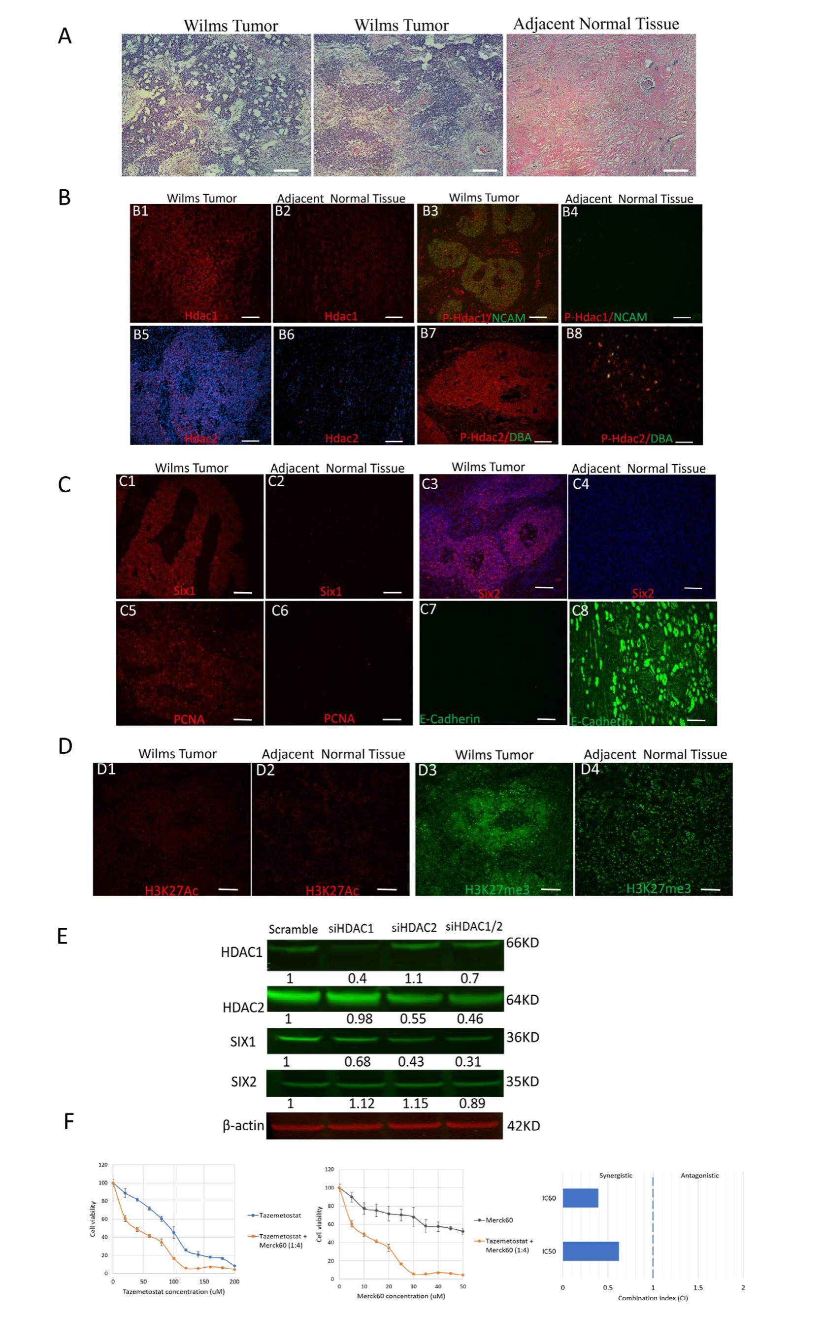
Wilms tumor (WT) is the most common childhood kidney cancer. Although WT is largely curable, current treatments fail in up to 15 percent of patients. Moreover, survivors suffer from the complications and late effects of the aggressive treatments. Thus, there is a critical need to improve our understanding of tumorigenesis to develop novel therapies to reduce the treatment burden while maintaining excellent survival rates. WT is believed to arise from the immature kidney cells, nephron progenitor cells (NPCs), which have failed to differentiate properly. Previous studies revealed that Wilms cells share a transcriptional and epigenetic landscape with normal renal stem cells. Although several studies have shown the positive associations between WT in children and embryonic exposure to adverse environments, the underlying mechanisms remain unknown. Altered epigenetics is central to oncogenesis in manypediatriccancers. The critical contribution of epigenetic dysregulation to pediatric tumors provides a compelling rationale for the therapeutic potential of epigeneticdrugs. Histonedeacetylases (HDACs) and Enhancer of Zeste Homolog 2 (EZH2, a histone H3K27 methyltransferase), have been demonstrated to play a critical role in self-renewal and differentiation of mouse NPCs. In addition, altered expression and mutations of HDACs and EZH2 have been linked to many human cancers, including WT. Thus, they are among the most promising therapeutic targets for cancer treatment. We reasoned that WT would result from the unrestrained proliferation of progenitor cells due to overactive HDAC1/2 (HDAC1 and HDAC2), and EZH2. We tested this hypothesis by analyzing a clinical specimen received from the left kidney tumor of an 11-year-old male patient diagnosed with WT and four other human WT specimens. We have the approval from Tulane Human Research Protection Office & Institutional Review Boards (Study number: 2019-623) to study WT specimens. The tumor (9.5 cm -8 cm -7.5 cm) is classified as favorable for the histology of indeterminate cell tumors. As shown in Figure 1A, the tumor exhibits a biphasic pattern with significant blastemal component admixed with stromal component. No significant epithelial component is present. The blastema represents the undifferentiated and malignant component, consisting of small round blue cells with overlapping nuclei and brisk mitotic activity. The blastemal component shows somewhat a basaloid growth pattern. The stromal component is also prominent and includes hypercellular undifferentiated mesenchymal cells.
