APE1 inhibition-promoted pyroptosis triggers T-cell infiltration and enhances anti-tumor immunity in NSCLC
APE1 inhibition-promoted pyroptosis triggers T-cell infiltration and enhances anti-tumor immunity in NSCLC
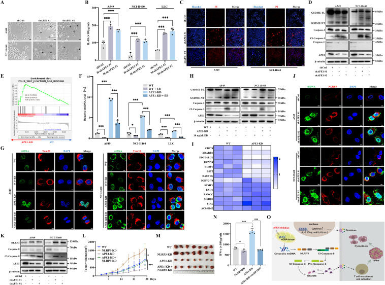
Pyroptosis, a form of pro-inflammatory programmed cell death mediated by gasdermin (GSDM) proteins, has been shown to synergize with immune checkpoint inhibitors to improve tumor eradication.1 The key regulators of pyroptosis are inflammasomes, particularly NOD-, LRR- and pyrin domain-containing protein 3 (NLRP3), which can be activated by cytoplasmic mitochondrial DNA.2 Our previous studies identified apurinic/apyrimidinic endonuclease 1 (APE1), a DNA repair protein, as a key factor in inducing pyroptosis in non-small cell lung cancer (NSCLC) upon its deficiency.3 However, the molecular mechanism by which APE1 inhibition leads to pyroptosis in NSCLC remains to be elucidated. To investigate the mechanism of APE1-mediated cell pyroptosis, we used specific shRNA to construct APE1 knockdown cells (hereafter referred to as APE1-KD) based on two human NSCLC cell lines (A549 and NCI-H460) and a murine LLC cell line (Fig. S1A and S1B). The indicative pyroptotic morphology was detected in these APE1-KD cells (Fig. 1A; Fig. S1C). It was found that the release of pro-inflammatory cytokines, including interleukin 18 (IL-18), interferon-gamma (IFN-γ), and IL-2, and chemokines, including C-C motif chemokine ligand 5 (CCL5) and C-X-C motif chemokine ligand 10 (CXCL10), was increased in the APE1-KD cells (Fig. 1B; Fig. S1D–S1H). Additionally, the lactate dehydrogenase (LDH) level was higher in the APE1-KD groups than in the control group (Fig. S1I). The APE1-KD tumor cells also showed an incomplete cell membrane as indicated by Hoechst/propidium iodide staining (Fig. 1C; Fig. S1J). Mechanistically, APE1 depletion specifically activated the caspase-3–GSDME pyroptosis pathway, without affecting GSDMA, GSDMB, GSDMC, GSDMD, or caspase-1 (Fig. 1D; Fig. S1K and S1L). To further explore the mechanism by which APE1 depletion leads to pyroptosis in NSCLC cells, RNA sequencing (GEO: GSE220807) was performed to investigate the potentially regulated signaling pathway. Gene Set Enrichment Analysis (GSEA) revealed that the pathways associated with DNA binding were enriched in the APE1-KD cells (Fig. 1E). Consistently, the APE1-KD tumor cells showed higher levels of cytoplasmic dsDNA compared with the wild-type (WT) cells (Fig. S2A). Previous studies have identified that APE1 inhibition leads to an increase in the amount of mtDNA copy number in tumor cells.4 Using quantitative PCR to detect mtDNA levels by targeting the D-loop region (the origin of mtDNA replication), we found that the expression of cytoplasmic mtDNA in the APE1 depletion cells was higher than that in the control cells (Fig. S2B). To determine whether APE1 inhibition promotes pyroptosis through cytoplasmic mtDNA accumulation, we depleted mtDNA, which resulted in reduced caspase-3 activation and GSDME cleavage (Fig. 1F–H; Fig. S2C and S2D).