Drosophila phenylketonuria modeling helps reveal the disease etiology and the modulation role of iron
Drosophila phenylketonuria modeling helps reveal the disease etiology and the modulation role of iron
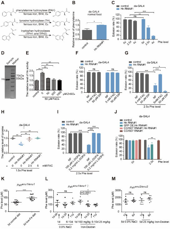
Phenylketonuria (PKU), one of the most common genetic metabolic disorders, is due to defective activity of phenylalanine hydroxylase (PAH), a member of the aromatic amino acid hydroxylases. In PKU patients, phenylalanine (Phe) levels in the blood and brain increase with clinical manifestations of severe intellectual disability and some other abnormalities. The exact mechanism and impacting factors of PKU are not completely elucidated. The therapy for PKU is mainly through a Phe-restricted diet to limit the uptake of Phe. In addition, tetrahydrobiopterin serves as an adjuvant pharmacologic therapy, primarily by its possible activity-boosting effect as a cofactor for PAH. Besides tetrahydrobiopterin, it is known that PAH additionally requires Fe2+ and O2 as cofactors (Fig. 1A). In this work, we generated a D. melanogaster PKU model to explore its regulatory mechanism. We found that the Phe-sensitivity of PKU Drosophila was strongly modulated by iron, and the eclosion defect could be almost completely rescued by tyrosine and L-3,4-dihydroxyphenylalanine (l-DOPA). The effect of iron on PKU was further confirmed in mouse PKU models.