Proteomic profiling of exosomes leads to the identification of a candidate biomarker for prostate cancer progression
Proteomic profiling of exosomes leads to the identification of a candidate biomarker for prostate cancer progression
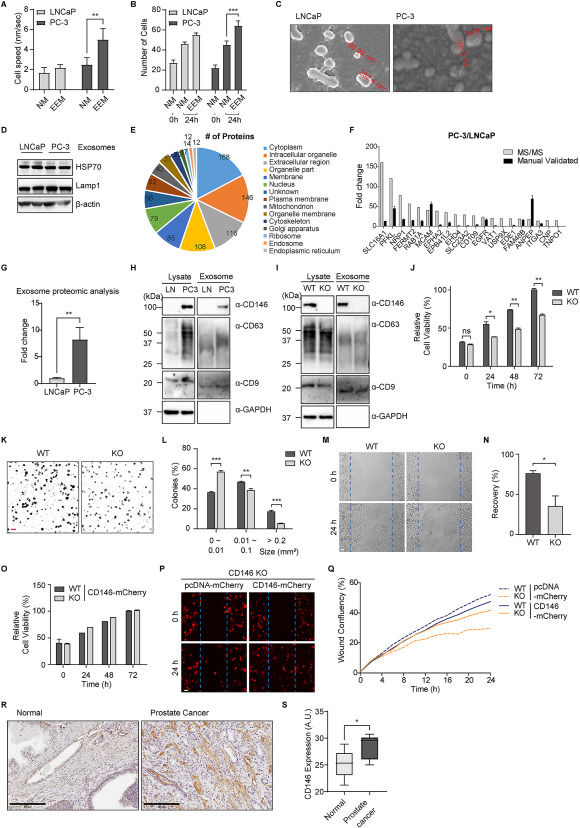
Extracting exosomes from bodily fluids offers a promising approach to overcoming inherent limitations, enabling the identification of potential biomarkers, especially those present in low abundance.1 PC-3 cells, known for their high metastatic potential compared with LNCaP cells, are widely used as a model for prostate cancer progression.2,3 However, the mechanisms underlying their metastatic characteristics remain unclear. Therefore, there is a critical need to investigate diagnostic methods for prostate cancer, as current techniques have various limitations that can lead to overtreatment due to their lack of precision.3 This study isolated exosomes from PC-3 and LNCaP cells through sequential ultracentrifugation to analyze their proteomic profiles using nano-liquid chromatography coupled to tandem mass spectrometry. Western blotting and quantitative reverse transcription-PCR validation revealed a higher abundance of CD146 (MCAM) in PC-3 exosomes, indicating that aggressive prostate cancer exhibits elevated levels of adhesion or cohesion proteins.4 We demonstrated that knocking out CD146 in PC-3 cells via the CRISPR-Cas9 system inhibited cell proliferation and invasion. Overall, CD146 was strongly associated with PC-3 cell mobility in vitro, suggesting that CD146 is a valuable candidate biomarker for prostate cancer progression.