Factor analysis and subtyping significance of CTNNB1 gene mutation detection in adamantinomatous craniopharyngioma
Factor analysis and subtyping significance of CTNNB1 gene mutation detection in adamantinomatous craniopharyngioma
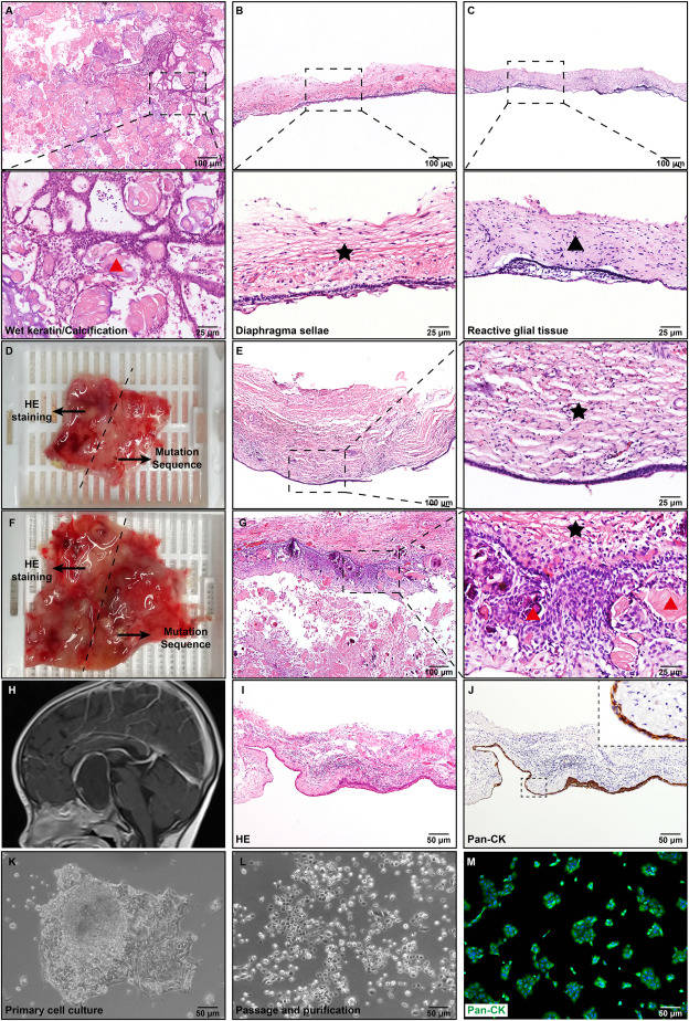
Craniopharyngioma (CP) is a rare, histologically benign tumor located in the sellar region which is defined as a grade I tumor by the World Health Organization (WHO) classification.1 There are mainly two different clinicopathological subtypes of CP, the adamantinomatous CP (ACP) and the papillary CP (PCP).1 Although both variations have distinct histomorphological characteristics, an accurate diagnosis might be difficult to make, especially in tiny and/or fragmented specimens. Furthermore, there is a continuous scientific dispute about the occurrence of mixed forms and the cell of origin of these tumors.2 The CTNNB1 gene mutation has been demonstrated to play a significant role in the tumorigenesis of ACP, and a growing body of research supported a high prevalence of CTNNB1 mutation in ACP, while many studies have failed to detect the CTNNB1 mutation in some ACP samples, thus resulting in a CTNNB1 mutation rate of 16%–100%.3,4 Previously, work by Apps et al (2020) has demonstrated the high prevalence of CTNNB1 mutations in ACP (100% in this study) when using a more sensitive TAm-seq sequencing method rather than Sanger sequencing.5 All the 22 ACP samples analyzed were found to carry the CTNNB1 mutation by TAm-seq. A low mutant allelic frequency was found to correlate with the failure to detect the CTNNB1 mutation by Sanger sequencing. Here, to figure out why the mutation rate of CTNNB1 in ACP was inconsistent in the literature and to highlight the importance of the mutation for CP subtyping, Sanger sequencing was used to detect CTNNB1 mutation in fresh-frozen tissues, formalin-fixed paraffin-embedded (FFPE) tissues, and primary ACP cells. Briefly, we observed that the CTNNB1 mutation detection was influenced by the wet keratin/calcification, diaphragma sellae, and reactive glial tissue in ACP tissues. Hematoxylin and eosin (H&E) staining can be used to guide mutation identification to enhance the rate of CTNNB1 mutation detection. An alternative for improving the mutation detection rate is to use primary ACP cells. Finally, The CTNNB1 mutation is critical for CP subtyping, particularly for atypical CP.